The annual Alzheimer’s Association International Conference (AAIC) took place earlier this summer in Amsterdam, the Netherlands. The conference brings together experts from around the world to share the latest advancements in Alzheimer’s disease and related dementias.
This blog post summarises the research presented by our team at AAIC and covers a variety of topics such as novel biomarkers, digitalised testing, disease staging measures, advances in neuroimaging, as well as patient engagement.
Biomarkers
Sophie Goldsmith – Optimising remote sampling of NfL and GFAP using blood cards
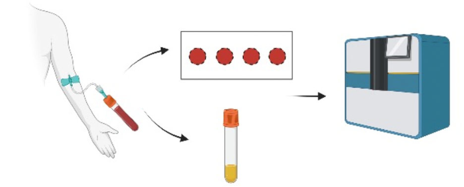
Remote collection of blood samples via methods such as blood cards would offer a more accessible and cost-effective alternative to standard clinic visits, whilst allowing more frequent samples to be collected. Sophie’s pilot study work considered the use of blood cards in the remote collection of neurofilament light chain (NfL) and glial fibrillary acidic protein (GFAP). To do so, she compared the concentrations of GFAP and NfL found in blood plasma samples versus in whole blood pipetted onto a blood card. She found strong correlations between GFAP concentrations in the plasma and whole blood card samples, however, the same was not true for NfL concentrations. These results suggest that GFAP concentrations can be measured successfully using remote collection via blood cards.
To read Sophie’s poster, see here.
Aitana Sogorb-Esteve – Identifying synapse- and lysosome-related proteins in genetic frontotemporal dementia

Studying fluid biomarkers in genetic FTD is important for bettering our understanding of the underlying mechanisms of disease. Two of these disease mechanisms – synaptic and lysosomal dysfunction – were examined in Aitana’s work. She performed a targeted search of synapse- and lysosomal-related proteins in a dataset obtained from a study in cerebrospinal fluid samples, using a technique called Tandem Mass Tag (TMT). Aitana found two sets of synapse-related proteins that decreased in all symptomatic mutation carriers, whilst a set of lysosome-related proteins specifically decreased in symptomatic MAPT mutation carriers. These findings contribute important insights into the underlying disease mechanisms of genetic FTD and how they differ between genetic groups and across disease severity.
To read Aitana’s full poster, see here.
Imogen – Investigating new markers of disease in progranulin-frontotemporal dementia
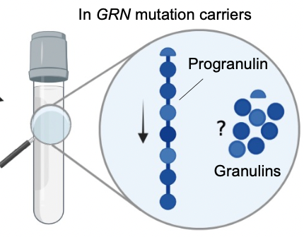
Mutations in the progranulin (GRN) gene lead to decreased levels of the progranulin protein in bodily fluids (such as blood and spinal fluid) and are a key cause of FTD. This progranulin protein can be broken down in 7 granulins and 1 paragranulin peptide and these subunits likely have key roles in FTD pathology. Imogen investigated whether these subunits could be measured in spinal fluid. To do so, she developed a panel that targets granulin 6, granulin 7, and paragranulin, finding that the concentration in the spinal fluid of each of these were significantly reduced in GRN mutation carriers compared to non-carriers. Imogen’s results contribute important findings to the underlying picture of GRN mutations and their role in disease. Furthermore, her work could be implemented in future clinical trials, serving as a novel biomarkers to see if therapeutic drugs can restore levels of granulins in the body’s fluids.
To see more of Imogen’s study, see here.
Clinical
Phoebe – Examining changes in disease severity over time in genetic frontotemporal dementia

The CDR®+NACC FTLD is a well established measure of disease severity across the frontotemporal dementia (FTD) spectrum, but few studies have assessed longitudinal changes in scores in genetic FTD. Phoebe’s work investigated changes in CDR®+NACC FTLD disease severity scores from participants’ first and second GENFI visit, across the three main genetic groups (chromosome 9 open reading frame 72 (C9orf72), progranulin (GRN), and microtubule associated protein tau (MAPT)). The greatest change in disease severity scores were found in the GRN mutation carriers, followed by C9orf72 expansion carriers, with MAPT mutation carriers showing the least. Her results also indicated that greater change was seen between first and second visit the more severe disease was. These findings therefore confirm the CDR®+NACC FTLD can measure changes in disease progression across FTD genetic mutation groups.
For Phoebe’s full study, see here.
Lucy – Using participant engagement to inform clinical trials and observational research
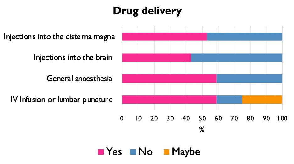
Lucy explored what types of clinical trials and treatments individuals with familial FTD would like to participate in. She did so using a questionnaire that considered people’s willingness to participate in trials and their knowledge about proximity to symptom onset, as well as the delivery of a study drug and their thoughts on gene therapies. She found that only 6% of individuals with familial FTD would not want to participate in a clinical trial, with the majority of people willing to: travel for a trial, find out their NfL levels, and uncover whether they were approaching symptom onset. Overall, Lucy’s work emphasises the importance of participant engagement in designing future observational research and clinical trials.
To read more on Lucy’s findings, see here.
Eve – Using the frontotemporal dementia rating scale as a measure of disease trajectory
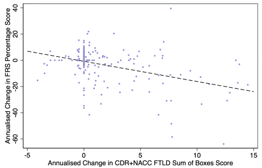
The FTD Rating Scale (FRS) is a sensitive measure of disease severity, though its use over time as a measure of disease severity in genetic FTD is less well understood. Eve’s study compared the change in score on the FRS at a baseline (first) GENFI visit and follow-up visit, across the genetic groups (C9orf72 expansion carriers, MAPT mutation carriers, and GRN mutation carriers). Eve found that as disease severity progressed, greater change in FRS scores were seen across the genetic groups. Also, changes in FRS scores were closely related to the changes seen between these timepoints (baseline and follow-up) on two other measures of disease severity (the MMSE and the CDR®+NACC-FTLD-SB), suggesting the FRS is a sensitive measure of disease.
See Eve’s full work here.
Digital Testing
Kerala – Examining practice effects in computerised cognitive testing

Practice effects refer to improvements in performance that occur when individuals undergo repeated testing. There is growing evidence that a reduction in practice effects can indicate early changes in cognition. Kerala’s study explored how practice effects differ when participants undergo ‘burst testing’ (baseline testing, 1 week, 2 weeks, and 4 weeks) using an iPad-based set of 16 cognitive tests called Ignite. She found significant practice effects across the timepoints in the healthy controls and presymptomatic individuals (improvement on 11/16 and 13/16 cognitive tests, respectively), with symptomatic individuals demonstrating significant practice effects on only 3/16 tests. Additionally, the presymptomatic group showed less of a practice effect compared to the healthy controls on several tasks. Her results suggest that practice effects may be diminished in people with symptoms, and that a decrease in practice effects could be an early sign of cognitive impairment in presymptomatic carriers.
Read Kerala’s full work here.
Rhian – Assessing cognitive impairment in frontotemporal dementia using a portable eye-tracking device
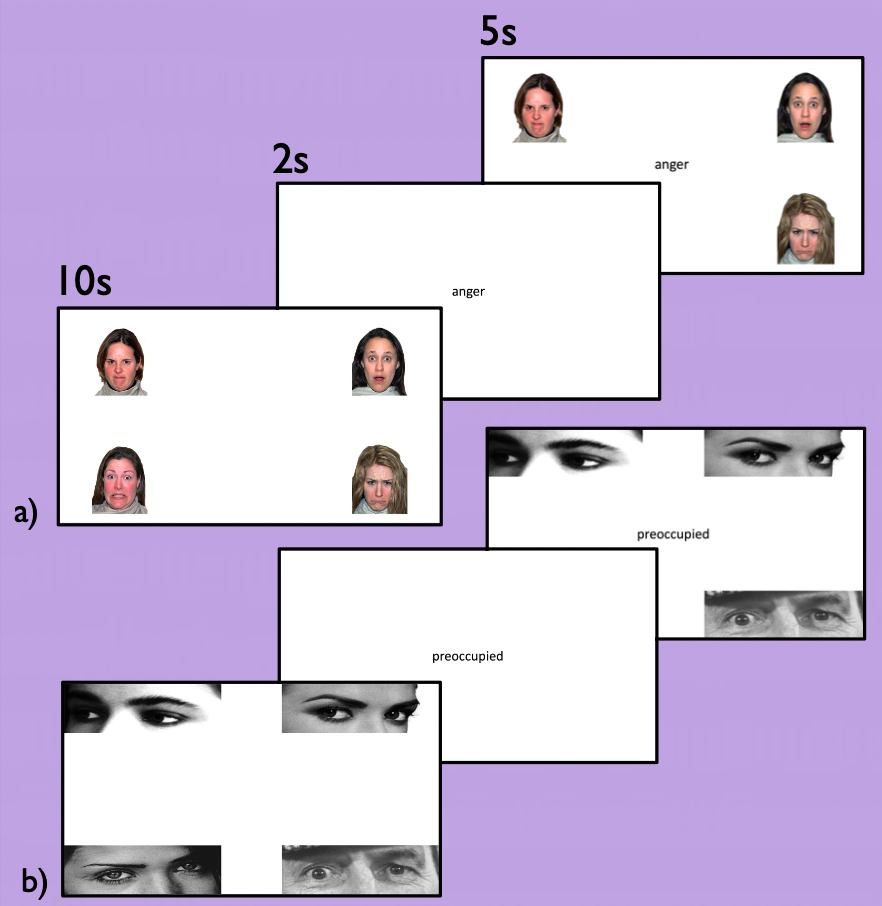
Rhian developed a portable eye-tracking protocol to investigate impairment in social cognition in individuals with presymptomatic FTD. To do so, presymptomatic mutation carriers and non-carriers were assessed on a task of simple and complex emotion recognition. Her results showed that presymptomatic mutation carriers had difficulty correctly identifying the target emotion, with reduced gaze on the target image compared to non-carriers. Her study suggests that the portable eye-tracking device has the potential to serve as a low-cost biomarker for monitoring disease progression in clinical trials, both at home and in clinical settings.
To read Rhian’s full poster, see here.
Imaging
Mica – Using inflammatory and synaptic PET imaging in individuals with C9orf72-frontotemporal dementia

Neuroinflammation and synaptic dysfunction are key changes seen in neurodegenerative diseases such as FTD. Mica’s work assessed both these changes in three C9orf72 expansion carriers with FTD (C9orf72-FTD) using PET imaging. Specifically, a PET tracer called PBR28 was used to investigate neuroinflammation and UCB-J was used to examine synaptic dysfunction. The images revealed significant neuroinflammation and synaptic dysfunction in the C9orf72-FTD participants using these tracers. When the PET results were compared to structural MRI data, she found that changes in both PET scans from patients compared to healthy controls were greater than changes in MRI scans, suggesting both tracers might provide more sensitive markers of disease. Of note, there was variability in PET tracer signal between the participants, with greater differences in UCB-J than PBR28 at earlier stages of disease. This implies that synaptic dysfunction may precede neuroinflammation and that disease duration may explain some of the variability in PET signal between individuals. Future work will explore the same markers of disease in larger cohorts.
Read Mica’s full study here.
Martina – Investigating white matter structural changes in genetic and non-genetic frontotemporal dementia

Although FTD is characterised by changes to brain tissue, such as white matter integrity, these alterations have yet to be investigated across different forms of FTD using a new technique called neurite orientation dispersion and density imaging (NODDI). So, Martina’s work used these NODDI sequences to extract two measures of white matter from brain scan images – the neurite density index (NDI) and the orientation dispersion index (ODI) – and compared these indices between symptomatic individuals with genetic and non-genetic (sporadic) forms of FTD. Abnormal values on the NDI and ODI were found across FTD diagnoses, with sporadic forms of FTD demonstrating more extensive and widespread changes in white matter compared with their genetic counterparts. Overall, her comparison of the NDI and ODI measures implies that the changes in white matter observed in individuals with symptomatic FTD are primarily related to a reduction in the density of neuronal axons (as measured by the NDI).
Read Martina’s full study here.
Phoebe, on behalf of the FTD talk team.
Keep up to date on all things research by following us on twitter – @FTDtalk / @GENFI1